


Calcium obtained from the diet is absorbed into the body and plays a vital role in building strong and flexible bones. Professor Ritsuko Masuyama developed genetically modified mice lacking the action of vitamin D, which has long been considered essential for calcium absorption, and she has identified a pathway in which the intestine independently regulates calcium absorption in response to phosphorus intake, revealing a new mechanism for calcium absorption that does not rely on the action of vitamin D.
Vitamin D maintains calcium levels and supports bone health
“We are what we eat.” This familiar phrase is often used when discussing the importance of diet for health. Among the nutrients essential for maintaining the health of our bones, which form the foundation of our bodies, calcium and vitamin D are particularly important.
“Vitamin D promotes calcium absorption in the small intestine, helping to maintain stable calcium levels in the body and supporting bone health. However, the detailed mechanisms of how vitamin D functions in different tissues remain mostly unclear,” explains Masuyama, who is working to unlock this mystery. To elucidate the mechanisms of vitamin D, Masuyama has developed vitamin D receptor (VDR) knockout mice in which vitamin D activity is selectively eliminated in specific tissues.
“The mechanism by which calcium is absorbed in the small intestine through the action of vitamin D has long been studied as a pathway for transporting calcium across cell membranes,” Masuyama says, explaining this process as follows (Fig. 1, right side).
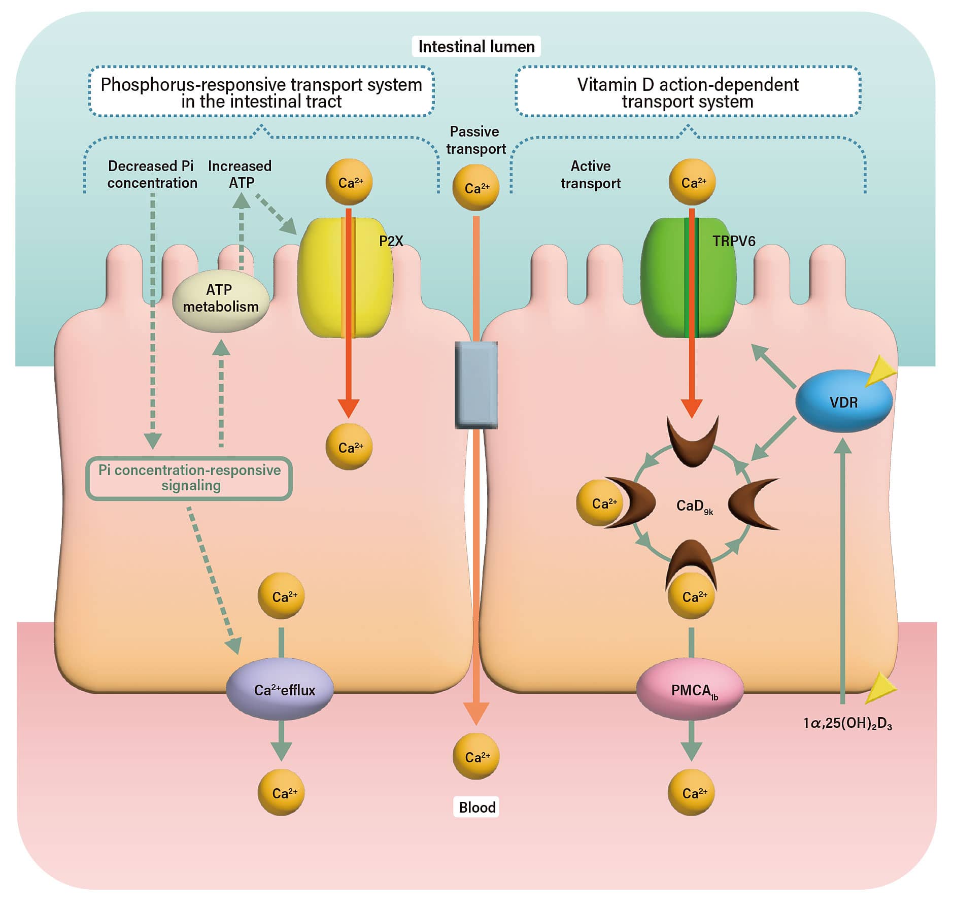
Dietary calcium enters intestinal epithelial cells from the lumen through calcium channels (TRPV6) expressed on the cell surface. Next, intracellular calcium ions are rapidly captured by calcium-binding proteins (e.g. CaD9k) and transferred to the calcium pump (PMCA) localized on the basolateral side. There, using the energy of adenosine triphosphate (ATP), calcium is actively pumped out of the epithelial cells into the bloodstream, completing the absorption process.
A key factor in this sequence of processes is the active form of vitamin D, or 1,25(OH)₂D₃. “When 1,25(OH)₂D₃ binds to the vitamin D receptor (VDR), it strongly promotes the expression of TRPV6 and CaD9k, both of which are essential for calcium absorption,” Masuyama notes. In her small intestine-specific VDR knockout mice, the reduced action of vitamin D leads to decreased expression of these proteins, resulting in a marked impairment of calcium absorption. In other words, vitamin D supports the entire process by which dietary calcium passes through cells of the small intestine into the bloodstream.
When calcium levels are high, vitamin D inhibits bone mineralization
Does increasing vitamin D intake simply lead to greater calcium absorption and stronger bones? According to Masuyama, this is a common misconception. “Vitamin D is often misunderstood to increase bone mass, but in fact it functions to maintain calcium homeostasis in the blood. It does not ‘increase’ bone, so to speak. Moreover, the mechanism by which vitamin D enhances calcium absorption becomes more effective when dietary calcium intake is insufficient,” she explains. In her experiments, when mice are given calcium-rich diets or when calcium is abundant in the gastrointestinal tract, the expression of the intestinal epithelial calcium channel (TRPV6) decreases. Despite this, calcium absorption is maintained at appropriate levels, indicating the presence of an alternative absorption mechanism.
The belief that vitamin D plays a role in maintaining calcium homeostasis but does not promote bone formation has also been confirmed from experiments using cultured cells. When 1,25(OH)₂D₃ was applied to osteoblast precursor cells, the resulting VDR signaling was found to inhibit mineralization around osteoblasts.
A newly discovered mechanism whereby limiting phosphorus intake enhances calcium absorption
Masuyama has also identified a novel pathway that promotes calcium absorption, in addition to the action of vitamin D.
“Reducing dietary phosphorus (Pi) intake can also improve the efficiency of calcium absorption,” she explains. In an experiment, VDR knockout mice that were fed a diet with normal calcium levels but reduced phosphorus intake (half that of a standard diet) showed increased calcium absorption and normalization of blood calcium levels, similar to the effects observed under high-calcium conditions.
To further investigate this mechanism, Masuyama recreated the intestinal lumen environment of the small intestine in an experimental system and examined how calcium absorption changes in response to phosphorus levels. This led to the discovery of a new regulatory pathway of calcium absorption involving changes in ATP metabolism within the gastrointestinal tract (Fig. 1, left side).
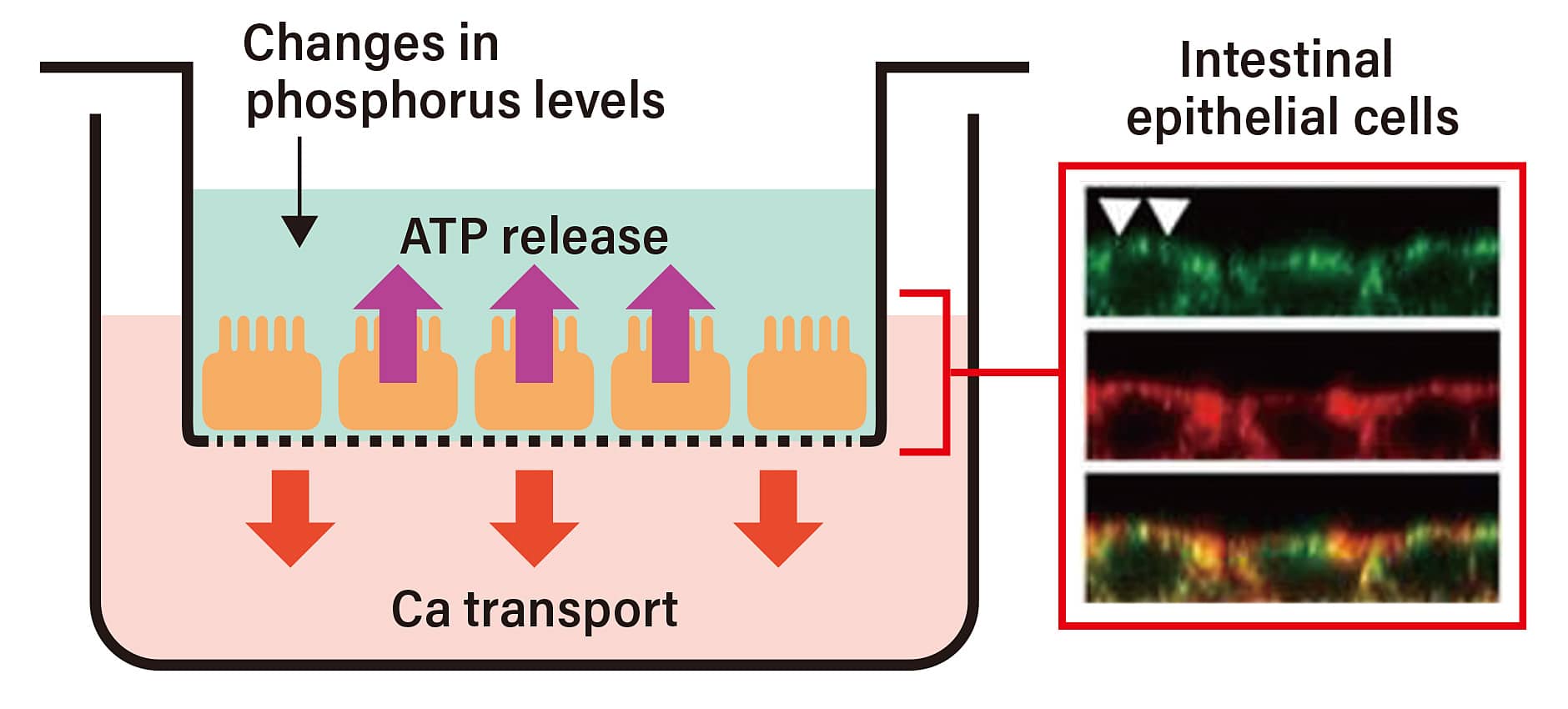
First, Masuyama cultured intestinal epithelial cells, induced their differentiation, and generated cells with polarity—specifically, a luminal side resembling the small intestine and a basal membrane side. Using these cultured cells, she analyzed how intracellular and extracellular conditions change in response to varying phosphorus concentrations. A key focus was the behavior of ATP. First of all, she found that when phosphorus levels are reduced, ATP release from cells into the intestinal lumen increases, while the expression of ENPP1—an enzyme that breaks down extracellular ATP—decreases.
Subsequent experiments showed that ATP binding to the ATP receptor (P2X) on the luminal membrane triggers calcium influx into the cells, followed by its release across the basolateral membrane into the bloodstream, effectively completing the absorption process. “These findings demonstrate that intestinal epithelial cells regulate calcium transport through changes in ATP metabolism in response to phosphorus levels in the gastrointestinal tract. In other words, independent of the action of vitamin D, the intestinal epithelium can detect changes in phosphorus levels and activate calcium absorption,” Masuyama explains.
Calcium absorption in the body is critically important not only for the growth of infants and toddlers, but also for the prevention of diseases such as rickets and osteoporosis. Further advances in Masuyama’s research are highly anticipated.